Screening and Diagnosing Flow Limitations in the Iliac Arteries
Clinician Information Document
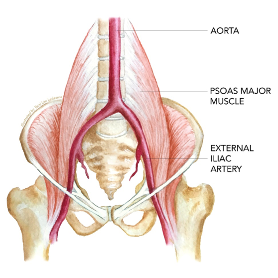
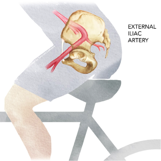
Images: Tami Lim Ledesma, anatamiart.net
Background
Blood flow limitations in the iliac arteries (FLIA), or external iliac artery endofibrosis (EIAE) is a sport-related vascular condition in which blood flow is limited unilaterally or bilaterally at the common (CIA) and/or external iliac arteries (EIA). Exercise capacity is prematurely impaired by ischaemia during vigorous exercise, causing claudication, pain, and powerlessness in the quadriceps and other working muscles.
FLIA typically affects young (< 50 yrs), otherwise healthy endurance-trained athletes, predominantly cyclists, speed skaters, and runners. Acute ischaemia is related to mechanical compression and kinking of the iliac artery during repetitive hip flexion in a bent-over (i.e. aerodynamic) racing position. Exposure to external mechanical compression and abnormal internal haemodynamic stress leads to pathological remodelling of the internal artery wall, called endofibrosis. Endofibrosis is histologically distinct from atherosclerosis or fibromuscular dysplasia.
There is no single gold-standard diagnostic criterion for FLIA. Clinical testing should be adapted to appropriately challenge the enhanced capabilities of this athletic population to ensure optimal diagnostic sensitivity. Diagnosis is interpreted based on a combination of assessment criteria as laid out in this document, synthesised from published literature and clinical experience.
The purpose of this document is to provide reproducible methodology for a non-invasive screening protocol for athletes suspected of having FLIA. The goal is to allow clinicians to perform this assessment outside of a specialised laboratory with commercially available equipment.
This document does not constitute personal medical or physiotherapy advice. A qualified clinician should be consulted in all cases of suspected sport-related vascular conditions.
Patient History
The investigation process for FLIA begins with a thorough patient history to understand the athlete’s current presentation and history of complaints. The patient’s training background, their injury history including previous treatments, and their medical history are valuable to understand contributing factors and consider differential diagnoses.
Vascular conditions are uncommon in endurance athletes. More common causes of non-specific complaints include musculoskeletal & orthopaedic sports injuries, biomechanical problems, issues with training load management, energy deficiency and nutrition limitations, cardiovascular concerns, and combined physiological & psychological stress from both training and non-training origin.
These differential diagnoses should be considered and assessed as part of the vascular investigation. Other sport-related vascular conditions to consider include popliteal artery entrapment syndrome (PAES) and chronic exertional compartment syndrome (CECS).
From the patient history, the following reports are suggestive of a flow limitation (Schep et al. 2002; INSITE Collaborators et al. 2016):
- Pain & claudication in the thigh or ≥3 muscle areas in the leg during exercise.
- Leg weakness, powerlessness.
- Resolution of symptoms quickly (within <5 minutes) after stopping provocative exercise.
- An absence of lower back problems (pain or mobility issues).
A questionnaire can be provided with discriminative questions for the patient to fill out.
Functional Biomechanical Assessment
There are not yet any specific, validated biomechanical assessment criteria for FLIA. Clinical experience suggests that examining the athlete’s general function off the bike, including mobility, stability, and strength of the thoracic and lumbar regions, hips, and pelvis can provide additional clinical information. However, the diagnostic sensitivity of functional tests needs to be established.
Athletes often present with transient, non-specific complaints with musculoskeletal / biomechanical concerns as an expected part of training and competing at a high level of sport. General physiotherapy / functional assessment can help to identify and treat these complaints. A neurological screen of the lower extremity should be performed to rule out red flags and consider referral for further testing.
Symptoms from a vascular origin may still show improvement with musculoskeletal / biomechanical management by relieving muscular compression on the arteries and reducing ischaemia, or by relieving secondary compensations occurring in the presence of ischaemia. However, it is currently poorly understood to what extent these components may be coincident with and contribute to FLIA.
Persistent complaints and insufficient response to musculoskeletal / biomechanical management may increase the index of suspicion for an underlying vascular origin.
From clinical experience, the following tests may provide clinical information:
- Standing trunk mobility (flexion, extension, rotation, side-bend).
- Standing squat (2-leg & 1-leg).
- Standing hip active range of motion (internal & external rotation, abduction, adduction).
- Seated slump test for neural tension.
- Neurological screen of lower extremity myotomes & dermatomes.
- Edge of bed Thomas test for rectus femoris & hip flexor.
- Supine resisted hip flexion (inner range with 90° hip & knee, outer range with straight leg).
- Supine FABER & FADDIR for hip joint mobility.
- Supine active hip rotation (internal & external, with 90° hip & knee).
- Side-lying hip abduction AROM & resisted.
- Prone hip & glute extension AROM & resisted.
- Prone elbow bridge straight leg extension AROM & resisted.
- 4-kneeling bird-dog contralateral & unilateral for anti-rotation stability.
- Trunk stability push-ups.
Other individual tests or a validated functional test such as the Functional Movement Screen (FMS) or Y-balance test may also be appropriate.
Provocative Exercise Test
A provocative exercise test should be performed to reproduce the athlete’s complaints, i.e. cycling or running. Cyclists can use their own bike on an electronically controlled trainer. The athlete should adopt an aggressive, hip-flexed racing position which will provoke symptoms for the highest detection sensitivity (where the absence of positive findings increases confidence to rule out the condition). Other sports can use cycling, treadmill running, or other modality-specific equipment.
An incremental exercise protocol to the maximal limits of tolerance is recommended. However, a custom protocol may be required to reproduce patient complaints (e.g. intermittent sprint efforts).
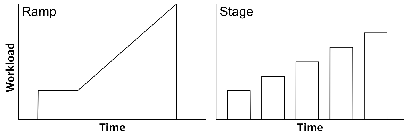
Typical cycling protocol is 4 min warm-up at 100 W or 1.0-1.5 W·kg-1, followed by a ramp rate equivalent to 20-40 W·min-1, depending on the athlete’s body size, sex, and fitness level, to achieve maximal task tolerance within 8-12 minutes. The ramp rate can be chosen to reach the athlete’s self-reported recent 5-min mean maximal power output within 10 minutes. The athlete may report their symptoms are more easily provoked with either shorter or longer test durations. Athlete feedback should be considered to inform the most appropriate test protocol.
Patients are instructed: “please report when you first begin to feel limited by symptoms”. The workload at this point is recorded along with location(s) and descriptions of symptoms. Patients may experience mild symptoms at low intensity or at rest. For this reason, careful wording as above is recommended to cue the patient to report when they feel their symptoms are the limiting factor to their performance. Onset of limiting symptoms is a useful outcome measure for longitudinally monitoring the effectiveness of management interventions.
Reporting pain and symptoms should be distinct from the perceived effort of exercise. After maximal exercise, the patient is asked to report both a rating of perceived exertion (RPE CR10 or Borg 6-20 scale) and a separate pain scale (CR10) to reflect the systemic effort and local pain, respectively (Borg 1998). Instructions for RPE: “Please report your maximum overall sensation of effort and fatigue during exercise from zero meaning no sensation of effort, to 10 meaning the most intense sensation of effort or difficulty ever experienced or imagined.” For pain: “Please report your maximum sensation of pain in your legs during exercise from zero meaning no sensations of pain, to 10 meaning the most intense sensations of pain ever experienced or imagined.”
Ankle-Brachial Pressure Index
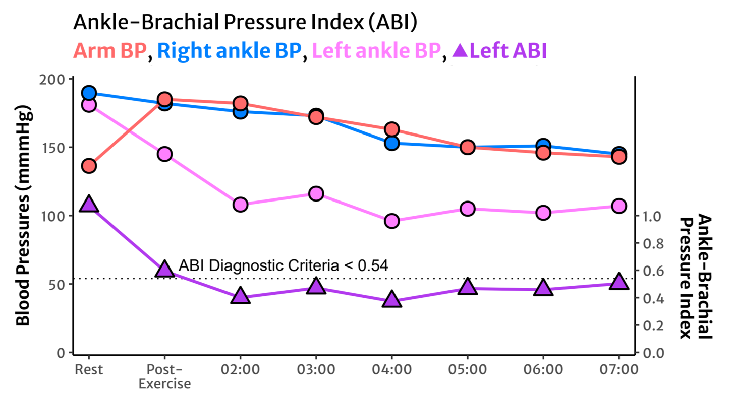
Blood pressure measurements should be taken bilaterally and simultaneously at the ankles, and unilaterally at the brachial artery for calculation of the ankle-brachial pressure index (ABI) before (resting baseline) and immediately (within 1 min) after exercise, with repeated measurements every minute or as frequently as possible for at least 5 minutes or until a return to baseline. Manual pressures can be taken alternating between ankles starting with the suspected unaffected/least affected leg. A handheld Doppler probe can assist with manual recording of ankle pressures.
ABI should be taken with the athlete remaining in the provocative (hip flexed) racing position with both legs in a symmetrical position, or rapidly moving to a seated position for greater screening sensitivity for a position-related kink. A stool or stable platform can be positioned quickly after exercise to symmetrically position both legs, as in Figure 3. Supine post-exercise ABI testing has higher diagnostic specificity (where the presence of positive findings increases confidence to rule in the condition) (Bender et al. 2004).
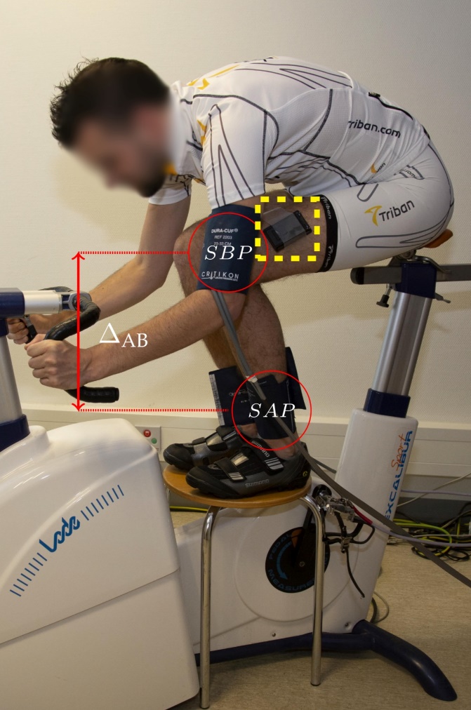
If ABI is taken in a seated position, the vertical distance from brachial artery to ankle should be measured and a correction factor used for ankle pressures (Gornik et al. 2008):
\[ ABI_{Seated} = \frac{SAP - (\Delta AB \cdot 0.78)}{SBP} \tag{1}\]
Where SAP and SBP are systolic ankle and brachial pressures, respectively, in mmHg. ΔAB is the vertical distance between ankle and arm, in cm. SAP is reduced by 0.78 mmHg for every 1 cm of vertical height between the arm and the ankle with the athlete in a seated position.
The following ABI criteria are suggestive of a flow limitation (Schep et al. 2002; INSITE Collaborators et al. 2016):
- Absolute ankle pressure <107 mmHg
- Bilateral ankle difference >23 mmHg
- ABI <0.54
Pedal Power Balance
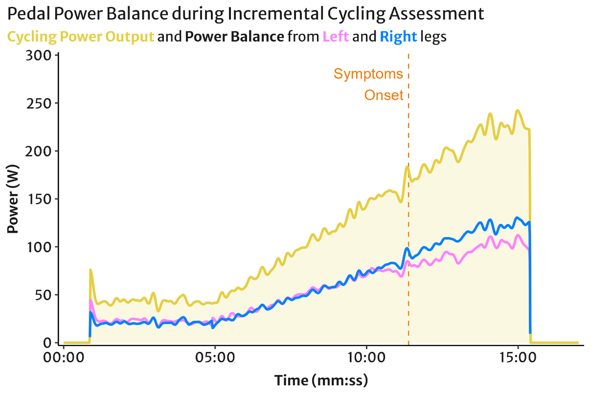
During a cycling test, a dual-sided power meter or cycle ergometer which can measure or estimate left/right power balance can be used to observe pedalling asymmetry.
Pedal power asymmetry is commonly observed in healthy athletes within a range of approximately 55/45% (in either direction), and can be greater in athletes with previous musculoskeletal injuries (Bini and Hume 2015). Thus, a power imbalance alone has low diagnostic specificity and is not suggestive of a flow limitation in isolation. However, when investigating a flow limitation, pedal power balance can be a helpful tool when combined with other diagnostic measures (Kleinloog et al. 2019).
The pattern of pedal power balance with increasing incremental workload may be more discriminative than an instantaneous absolute power balance. A ‘converging’ power balance trend is commonly seen in healthy athletes, where symmetry improves toward 50/50 with increasing workload.
A ‘diverging’ power balance trend is where power symmetry decreases away from 50/50 with increasing workload. A ‘cross-over’ trend is where one leg produces relatively greater power at lower workload, and less power at higher workloads. These latter two power balance trends may be more suggestive of pathology than the former, however diagnostic sensitivity/specificity is not yet established.
Near-Infrared Spectroscopy
Near-infrared spectroscopy (NIRS) devices are small, non-invasive, wearable devices which use optical measurements to estimate the saturation of oxygen (O2) on haemoglobin and myoglobin (muscle oxygen saturation; %SmO2). This provides information on the real-time balance of local muscle O2 delivery and uptake / consumption (Barstow 2019). SmO2 will decrease when O2 uptake is greater than delivery, e.g. deoxygenation during exercise. SmO2 will increase when O2 delivery is greater than uptake, e.g. reoxygenation during recovery. Delayed SmO2 reoxygenation after maximal provocative exercise is shown to improve diagnostic sensitivity for FLIA (van Hooff et al. 2022).
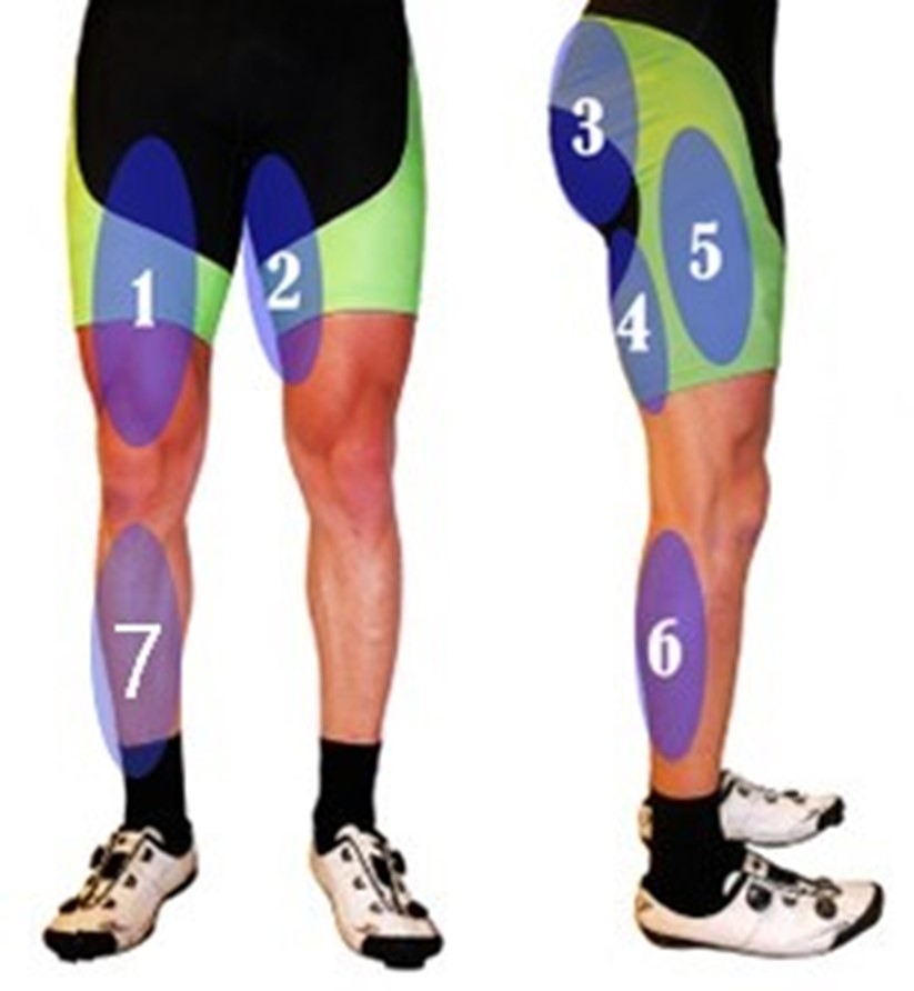
NIRS sensors are placed on bilateral quadriceps vastus lateralis (VL) muscle belly at ⅓ distance from the patella to the greater trochanter and secured with elastic tape or wrap, or by the athlete’s compressive elastic shorts. See Figure 3 above for approximate positioning. They can typically be recorded via Bluetooth to a cycling computer or one of several training apps.
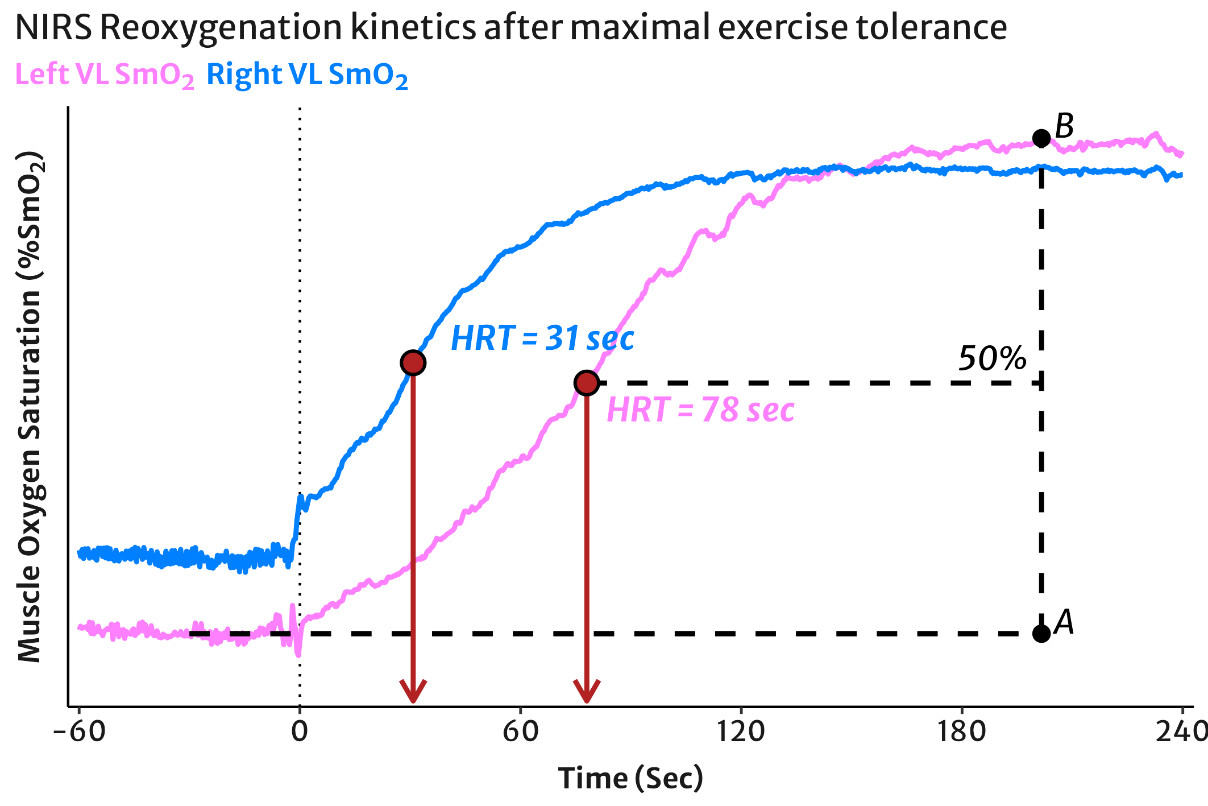
SmO2 reoxygenation time matters more than the SmO2 values themselves. Reoxygenation may be interpreted qualitatively by visually evaluating the time difference between the steepest segments of bilateral SmO2 responses at a scale of <5-min. A proportional time difference between SmO2 tracings as shown in Figure 6 below may be suggestive of a unilateral flow limitation.
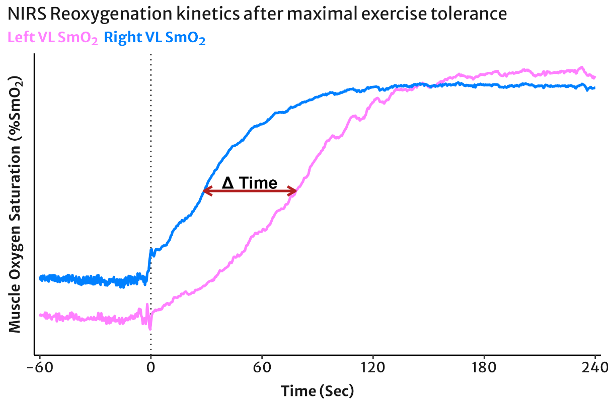
Quantitative NIRS data processing & analysis may improve diagnostic sensitivity. This can be performed in Excel or a programming language such as R or Python. A simple calculation can be done to quantify the half-recovery time (HRT). HRT is the time required after the end of maximal provocative exercise to resaturate half of the total SmO2 reoxygenation amplitude (van Hooff et al. 2022; Arnold, Yogev, et al. 2024).
\[ HRT\ (\text{sec}) = t \text{ when } SmO_2 = A + 0.5 \cdot (B - A) \tag{2}\]
Where HRT is equal to time (t) in seconds, and A & B are the starting and ending (e.g. peak) SmO2 values, respectively (see Figure 7). An open-source app is currently under development to make quantitative analysis more accessible to clinicians without coding requirements (mnirs: Muscle Near-Infrared Spectroscopy Data Processing and Analysis).
Diagnostic criteria are still being established for reoxygenation kinetics. Heuristics for screening assessment include: a difference >50% between legs may be suggestive of unilateral flow limitation. Absolute reoxygenation kinetics HRT >60 sec in endurance trained cyclists may be suggestive of unilateral or bilateral flow limitations (van Hooff et al. 2022).
Power-Deoxygenation Index
When both pedal power balance and bilateral NIRS muscle oxygenation are measured together, they can be used to calculate a combined metric called the power-deoxygenation (PD) index (Arnold et al. 2022). The PD-index is calculated as the ratio of unilateral power output relative to NIRS deoxygenation. This can be interpreted as a relative scale of the metabolic disruption occurring at a given power output. Or as a gross proxy for internal metabolic load relative to external load. In the most simple equation, the area above the SmO2 signal is used to quantify deoxygenation:
\[ PD\ (\text{a.u.}) = \frac{\text{power output (W)}}{100 - SmO_2\ (\%)} \tag{3}\]
The PD-index is useful to suggest whether a limitation is occurring either on the muscle recruitment (demand) side, or the blood flow and O2 delivery (supply) side. If one leg is producing less power and has a higher PD-index, it suggests less metabolic disruption is occurring and the limitation may be more on the demand side, pointing toward a musculoskeletal limitation. If the leg has a lower PD-index, this may suggest that O2 delivery is the limiting factor to power output, suggestive of a flow limitation. Like pedal power balance, the bilateral trend with increasing workload may be more meaningful than any one absolute difference.
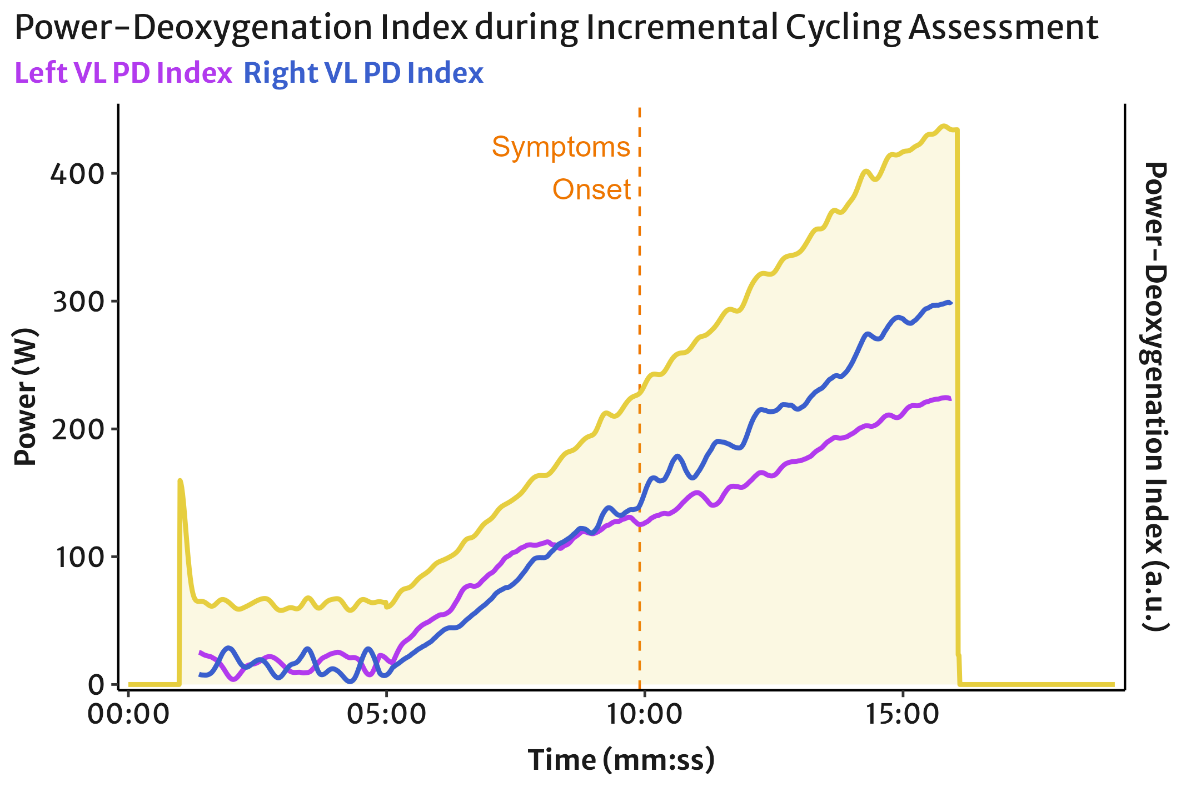
The relative nature of this metric means that comparisons are only valid during a single exercise trial within a single patient. PD-index values are particularly sensitive to baseline tissue differences under the NIRS probe and the accuracy of the power meter and pedal power balance recording. Thus, normative values for the PD-index cannot yet be provided and further research is required. PD-index should be interpreted with caution alongside other diagnostic measures.
Advanced Diagnostic Imaging
When FLIA is ruled in based on functional and provocative exercise testing as above, referral is recommended to a vascular specialist clinic for advanced diagnostic imaging. Imaging methods include echo-Doppler (duplex) ultrasound, intravascular ultrasound, magnetic resonance angiography, and/or computed tomography scan.
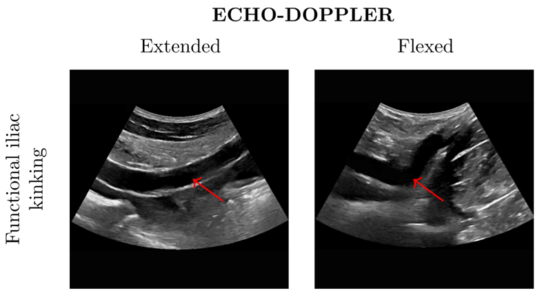
Scans should ideally be performed in the provocative hip flexed position in addition to the standard supine position, to improve detection sensitivity for arterial kinking and tortuosity (Schep et al. 2001; Peach et al. 2012). Quantitative imaging values (such as arterial diameters) should be interpreted with consideration of the patients’ fitness level and training history, rather than relying on population-based standards.
Standard clinical criteria may be established from a much different population, e.g., untrained individuals or vascular disease patients. For example, highly endurance-trained athletes are expected to have larger arterial diameters than population-predicted normative values would suggest (Green et al. 2012). Considering these characteristics will help improve diagnostic sensitivity when evaluating arterial stenosis from imaging.
Duplex ultrasound (DUS) is used to image the iliac arteries to detect kinking and measure peak systolic velocity (PSV) of blood flow through the aorta, CIA, and EIA bilaterally. Visualisations and PSV recordings are ideally taken at rest in 3 positions: (1) relaxed in supine hip extended, (2) relaxed in 90° hip and knee flexed position (e.g., with the patient’s leg resting on a stool), and (3) psoas-contracted in 90° hip and knee flexed (e.g., with the patient actively pushing against their knee to isometrically contract hip flexion). The hip flexed and psoas contracted positions are then repeated after the patient performs 50 standing squats to elevate blood flow. Imaging in a flexed-hip position may have higher diagnostic sensitivity with lower specificity compared to supine (D’Abate et al. 2017).
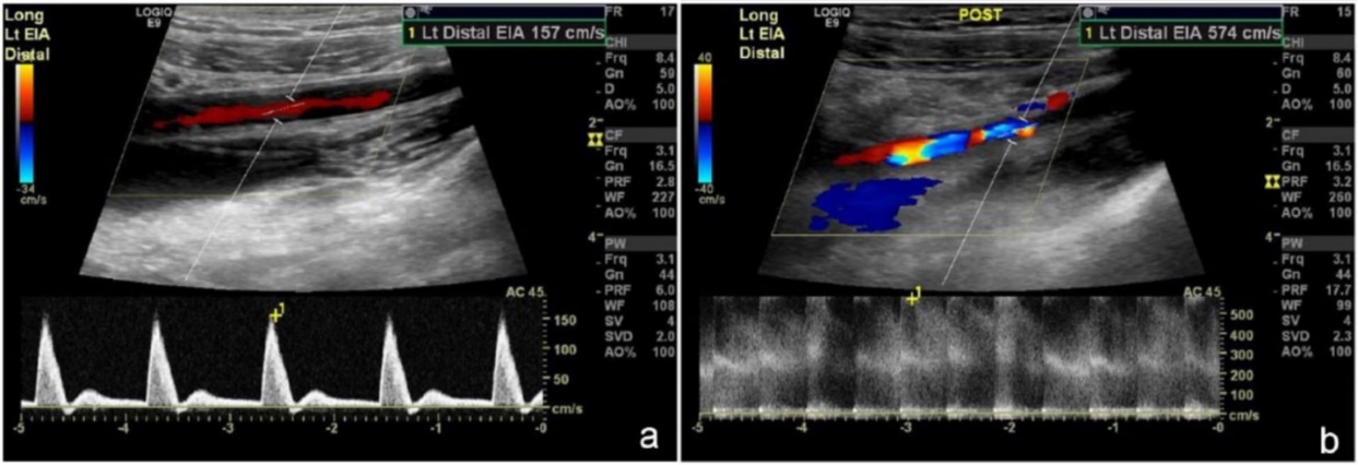
The following PSV criteria are suggestive of a flow limitation (Schep et al. 2002; Peake et al. 2018):
- PSV hip extended > 1.48 m·sec-1
- PSV hip flexed > 1.70 m·sec-1
- PSV contracted > 1.70 m·sec-1
- PSV post-exercise > 3.18 m·sec-1
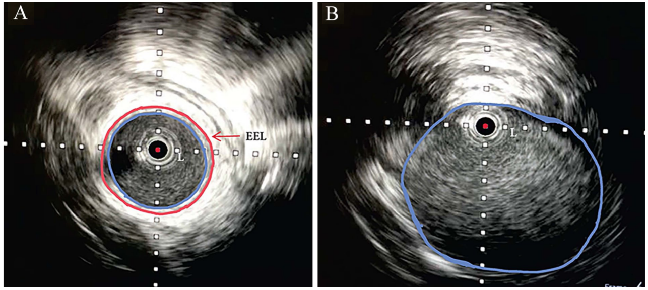
Intravascular ultrasound (IVUS) is becoming more common to evaluate arterial diameters and visualise the lumen for hypertrophy. Recently, a novel visual characteristic has been proposed to improve diagnostic sensitivity, called the ‘perfect circle appearance’; where the IVUS remains centred related to narrow and stiffened elastic lumen in the presence of endofibrosis (Sharifi et al. 2024).
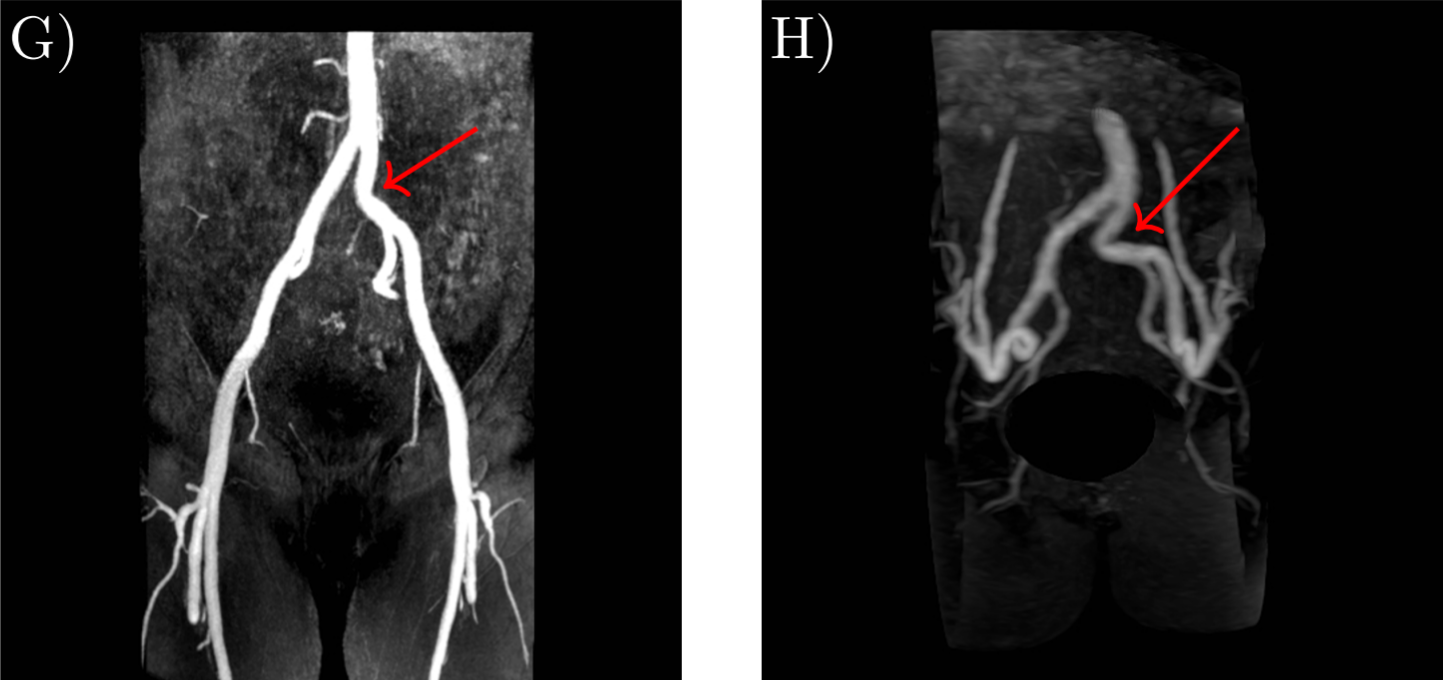
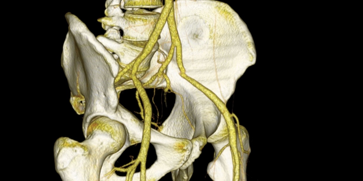
Magnetic-resonance angiography (MRA), digital subtraction angiography (DSA), or computed tomography (CT) can be used to visualise the complex, three-dimensional vessel geometries in the pelvis region for pre-operative surgical planning. 3D reconstruction will help to localise the lesion and observe tortuosity outside of the limited viewing plane of ultrasound.
Imaging in both supine and hip-flexed position is ideal for diagnostic sensitivity and specificity (Schep et al. 2001; Peach et al. 2012). Athletes can be positioned on their side with thighs flexed, or in crook position with a wedge under the thighs, within the limits of the scanner bore.
Endofibrosis may not be associated with detectable artery wall thickening, therefore evaluation of lumen diameter is the primary outcome (Perrier et al. 2020). Even “mild” reduced artery diameters (e.g. by 20%) observed at rest, which may be sub-clinical in a sedentary population, may contribute to severe symptoms during the greatly elevated demands of exercise (Rouvière et al. 2014).
Thus, interpretation of arterial diameters should consider contralateral narrowing where appropriate, and interpret absolute diameters with reference to the body size, sex, and training status of the patient (Green et al. 2012).
Conservative Management
The most common recommendations for reducing symptoms in FLIA are to reduce, modify, or stop participation in the provocative activity. When the provocative activity is stopped, the endofibrotic lesion is expected to stabilise and symptoms are often reduced or eliminated (Abraham et al. 1997). However, the advice to entirely stop sport is often unsatisfactory to athletes and additional treatment options should be considered (Arnold, Mawji, et al. 2024).
Surgical reconstruction is the definitive treatment for endofibrosis (INSITE Collaborators et al. 2016). More information on surgical interventions and patient outcomes is available elsewhere (van Hooff et al. 2023) and should be discussed with an experienced vascular surgeon. Conservative management options can be implemented during the investigation process to help reduce symptom burden, improve the patient’s functional fitness in preparation for eventual surgery, and most importantly as part of the rehabilitation and return-to-sport (RTS) process after surgery.
Symptoms do not have to be entirely eliminated during conservative management but can be controlled and minimised. The onset of performance-limiting symptoms should be considered as the athlete’s functional training threshold, even if they are able to perform at a higher level by pushing to their maximal limit of symptom tolerance. The goal of conservative management is to first reduce symptoms to mitigate continued disease progression, and second to preserve performance within those limits.
Just like conservative management will not change the structure of the artery once endofibrosis has developed, surgical intervention may not address the underlying functional limitations which may result from and contribute to FLIA. Functional rehabilitation is an important part of a comprehensive treatment and RTS program. Planning RTS is an interdisciplinary collaboration involving the vascular surgeon, sports physicians, rehabilitation therapists, coaches, and the athlete themselves.
For cycling, a common recommendation is to modify bike fit to open the hip flexion angle and reduce the potential for compression and kinking of the iliac arteries (INSITE Collaborators et al. 2016). Bike fit modifications are highly individual and should be performed with an experienced clinician or bike fitter. In general, common adjustments can include:
- Raising the handlebars and using a shorter stem to reduce the athlete’s reach.
- Using a shorter crank to reduce hip flexion at the top of the pedal stroke.
- Adjusting the saddle fore-aft position and saddle tilt for comfortable pelvic rotation.
- Moving the shoe cleat rearward toward the mid-foot.
- Reduce actively pulling up on the pedals, even using shoes without clips if necessary.
Clinical experience suggests that spending the majority of training hours in a more upright body position may reduce symptom burden and preserve a higher fitness level, allowing athletes to race in their provocative position when required. Non-professional athletes can be encouraged to reduce provocative training volume and supplement their training with alternative activities such as running, skiing, strength training, and other exercises. Runners and other athletes can discuss with their coaches and clinicians about options for modifying training practices and body positions or muscle recruitment patterns to help minimise symptoms.
Athletes can be counselled to modify their training practices to help reduce symptom burden. For example, short intermittent intervals with frequent brief rests, and intervals distributed throughout a longer low-intensity training session can be used instead of a continuous high-intensity interval session. Performing intervals in a standing cycling position can help to re-distribute the metabolic load away from the symptomatic muscles while preserving the systemic adaptive training stimulus.
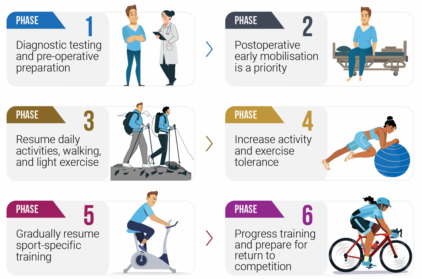
For patients who undergo surgical reconstruction, a graduated RTS process is critical to allow these athletes to regain their previous performance level. Surgical guidelines typically advise a 4-6-week rest period (e.g. maximum walking intensity) before re-introducing the provocative sport (INSITE Collaborators et al. 2016; Arnold, Mawji, et al. 2024).
It is common to continue to experience some symptoms when resuming progressive training. Symptoms will often continue to improve as perfusion within the working muscles is re-normalised, and the distal tissues remodel to the enhanced conduit artery blood flow.
Professional cyclists who have returned to sport following surgery for FLIA report that it can take 6-9 months to feel that they have regained their full performance (The Cyclists’ Alliance 2022). Some athletes will be able to return earlier, and unfortunately some athletes will not be able to return to their previous level at all without continued symptoms.
Patient & Clinician Considerations
FLIA is a condition which develops gradually and is difficult to detect and diagnose. For this reason, it is still not fully understood. Athletes, coaches, and clinicians should consider vascular conditions as a possible cause of unexplained performance impairment or exertional leg pain once more common diagnoses have been excluded or when response to initial treatment is inadequate.
Greater awareness of sport-related vascular conditions will improve clinical management; however, FLIA remains an uncommon condition, and should not be the first diagnosis for unexplained leg pain.
Performance decline may occur over months or years, and symptoms are often subtle or inconsistent. With invisible injuries such as concussion, overtraining, energy deficiency, or FLIA, identifying the source of limitation can be challenging for both the athlete and their support team, and it may be unclear whether a problem exists at all.
The experience of painful, unexplained loss of fitness can be a source of considerable distress to the athlete during what is often a prolonged, frustratingly slow investigation process. Athletes frequently report feeling that they are not believed or taken seriously by medical professionals, coaches, and other managers/support personnel. Whether intended or not, we as clinicians need to do a better job to ensure our athletes feel supported. Providing explicit trust, clear information, and consistent support can help reduce feelings of uncertainty and fear, and give the athlete greater autonomy during the diagnostic and management process, which itself is likely to improve outcomes.
Clinician Background
Jem Arnold is a physiotherapist and PhD researcher at the University of British Columbia. He has worked internationally with professional and amateur cyclists, runners, and other endurance athletes with sport-related vascular conditions. He uses his clinical, coaching, and research background to help athletes return to competition from injury, and to transition from competitive sport to sustainably training for lifelong fitness. As part of Jem’s PhD work, he has helped to develop screening tests for sport-related vascular conditions which are used internationally to improve patient management outcomes.

For consultation on athlete screening, please contact Jem: jem@jemarnold.ca
Screening Protocol Equipment
| Equipment | Examples |
|---|---|
| A stationary cycle ergometer or electronically controlled trainer to fit the athlete’s personal bicycle. Such as Tacx Neo or Wahoo Kickr. | 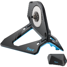 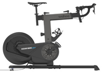 |
| A stationary cycle ergometer or power meter on the athlete’s personal bicycle, ideally capable of recording left/right pedal power balance. Such as Favero Assioma or 4iiii. | 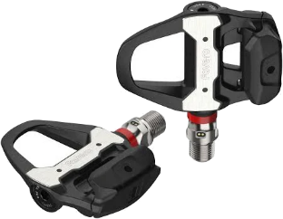 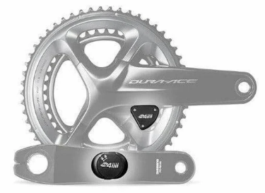 |
| A device or software to record cycling metrics (cycling power, power balance, cadence, heart rate, etc.). Such as Garmin Edge, PerfPro Studio Software, or Golden Cheetah. | 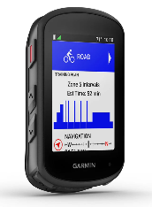 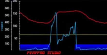 |
| 2x Near-infrared Spectroscopy devices for bilateral vastus lateralis muscle oxygenation. Such as Train.Red or Moxy. NIRS can be recorded to certain cycle computers, computer software, or mobile apps from the device manufacturer. |   |
| 3x Automatic or manual blood pressure recording devices for bilateral ankles & one arm. | 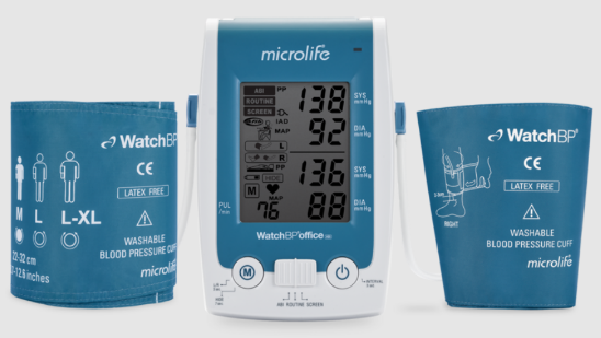 |
| Small supplies (elastic tape/wrap, goniometer, measuring tape, skinfold calliper, etc.) |