Sport-Related Vascular Conditions in Endurance Athletes
Flow limitations in the iliac arteries (FLIA)
Patient background information
Sport-related vascular conditions are uncommon but problematic conditions affecting the arteries of the legs in otherwise healthy, highly trained endurance athletes.
This page is general information, not medical advice. Discuss your symptoms with a registered clinician.
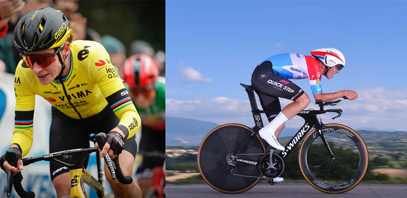
In recent years a rising number of professional cyclists and triathletes have been diagnosed with a debilitating vascular condition called blood flow limitations in the iliac arteries (FLIA). Marianne Vos (NED), Maggie Coles-Lyster (CAN), Sarah Gigante (AUS), Zdeněk Štybar (CZE), and Bob Jungels (LUX) are a few household names in the World Tour peloton who have publicly struggled with this condition.
Amateur cyclists can also develop FLIA. With less clinical knowledge available about vascular conditions in amateur athletes, detection and diagnosis is often a difficult and lengthy process.
Presentation
FLIA is an uncommon vascular condition where blood flow through the arteries of one or both legs is impaired during exercise.
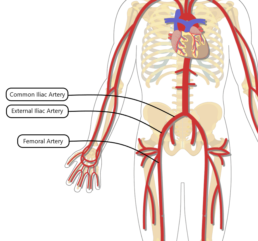
The iliac artery is a large blood vessel in the lower abdomen carrying blood from the heart to the legs. If flow through the iliac artery is limited, the leg muscles will not receive enough oxygen to meet the demands of exercise, causing symptoms, pain, and impaired performance.
During exercise the affected leg(s) may feel pain, burning, weakness, powerlessness, heaviness, cramping, tingling, and other “dead leg” sensations. Symptoms are usually worst during higher intensity or longer exercise and resolve within a few minutes of stopping. There are usually no symptoms at rest, although some patients notice symptoms during non-exercise activities such as climbing stairs.
Causes
The exact causes and risk factors for FLIA are not fully understood. The condition is thought to be related to the forward flexed racing position used in cycling and speed skating, combined with the repeated hip flexion and extension common to running and most endurance sports [1].
Not every cyclist or endurance athlete is at risk of developing FLIA. Suspected additional risk factors include previous injury around the hip, natural anatomic differences in the arteries, and changes in muscle activity with certain pedalling styles (see Treatment below).
Athletes may perform millions of hip flexions over years of training and competition. As the hip flexes toward the chest, the iliac artery can be compressed by surrounding tissues, particularly the repeatedly contracting iliopsoas (hip flexor) muscle. The artery can become kinked, like squeezing a garden hose to stop the flow of water.
Elite athletes can deliver 10-15+ litres of blood per minute to the legs during exercise. This high flow pushing through a kinked or compressed artery stresses the artery walls, which can abnormally thicken over time, a process called endofibrosis.
Over months or years of exposure the inside of the vessel narrows, further limiting blood flow during exercise and gradually worsening symptoms. FLIA and endofibrosis are like a chronic overuse injury. This process is different from common age-related cardiovascular diseases such as atherosclerosis or diabetes, and typically affects much younger and fitter individuals.
Prevalence
FLIA typically appears in relatively young, healthy endurance athletes under 50 years of any gender. It may be associated with longer training histories and higher lifetime training volumes. Its prevalence among professional and amateur athletes is unknown, but diagnoses are increasing as awareness is growing.
FLIA is uncommon and should not be the first suspected diagnosis for unexplained leg pain during exercise.
More common causes include orthopaedic injuries of the muscles and soft tissues around the hips and lower back, poorly managed training load, nutrition & energy imbalance, and training + non-training stress i.e. overtraining. Other sport-related vascular complaints to consider include popliteal artery entrapment syndrome and chronic exertional compartment syndrome, both common in runners.
Common orthopaedic conditions should be investigated and treated by a qualified clinician as part of the FLIA investigation. Treating these may improve symptoms whether or not a blood flow limitation is present. If the athlete does not respond to treatment as expected, FLIA or another vascular complaint may be suspected and investigated further. Multiple conditions can be present together.
Screening and Diagnosis
If FLIA is suspected, the first step is to review medical and training history to understand the presentation and history of complaints. A specialised questionnaire for detecting sport-related vascular problems can be provided to the athlete.
Next is a clinical assessment of the athlete’s movement and function, particularly of the lower back and hips.
This is followed by a provocative exercise test in which the athlete attempts to reproduce their symptoms. Provocative exercise should be cycling on a stationary bike or treadmill running, in the same body position in which the athlete experiences symptoms. An incremental ramp or multi-stage test with resistance increasing over 15-30 minutes is performed until the athlete reaches maximal effort or maximal tolerance of symptoms [2].
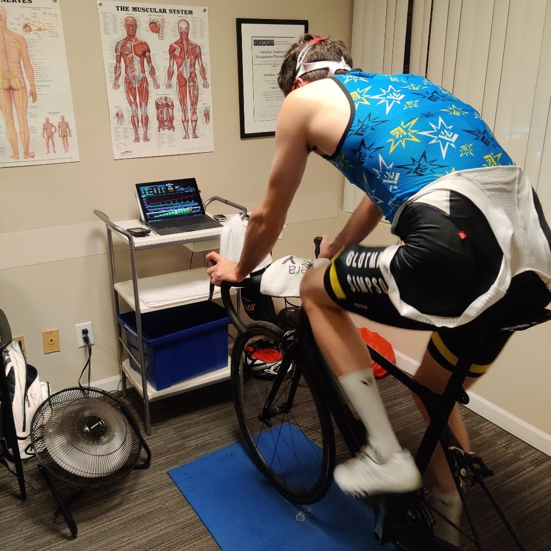
During and after the exercise test the clinician takes several measurements to help detect a blood flow limitation, including the workload at which the athlete first reports feeling limited by symptoms.
Near-infrared spectroscopy (NIRS) devices are small, non-invasive light sensors worn on the thigh muscles to monitor muscle oxygen saturation during exercise. The technology is similar to the optical heart rate sensor in a sport watch. These data can help differentiate a blood flow limitation from a muscle recruitment issue [3].
Immediately after the athlete reaches maximal exercise tolerance, blood pressure cuffs are placed around one arm and both ankles to measure the ankle-brachial index (ABI). The athlete either remains in the provocative cycling position or lies on their back with hips flexed toward the chest and knees supported on a stool [2].
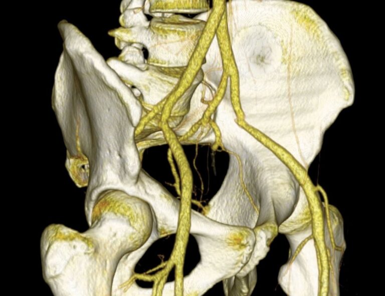
If these tests suggest FLIA, the athlete is referred to a sports physician or vascular specialist for further diagnostic evaluation. Echo-Doppler ultrasound images the arteries at rest and after a brief exercise task such as fifty standing squats. The left and right iliac arteries are imaged with the athlete lying on their back with straight legs, with knees flexed to the chest, and while pushing against their own knee to contract the hip flexors. This may reveal a kink, narrowing, or other abnormality in the artery [4].
Further diagnostic imaging may include peak systolic velocity with Echo-Doppler ultrasound, magnetic resonance angiography (MRA), computed tomography (CT), and/or intravascular ultrasound [5]. These provide more detail on artery structure and help guide treatment options such as surgical reconstruction.
Treatment
Options for conservatively treating FLIA are limited. The first step is to modify riding position and bike fit to reduce hip flexion: raising the handlebar, moving it closer to the body, and moving the saddle forward. Shorter cranks further open the hip angle. Focusing pedalling technique on pushing down rather than pulling up can reduce hip flexor contraction. These modifications can reduce symptom burden during training. Bike fit changes should be made with an experienced clinician or bike fitter [7].
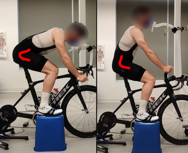
High intensity training can be reduced or modified to manage symptoms. Repeated short intervals with frequent brief rests can reduce symptoms while preserving fitness. Non-professional athletes may benefit from reducing provocative training and adding alternative activities. Clinician-guided rehabilitation and strengthening exercises can further reduce symptoms and maintain fitness.
In some cases conservative management is not sufficient. Athletes understandably may not want to give up their sport, and professionals rely on performance for their livelihood. For these individuals, surgical reconstruction can be considered.
Diagnostic imaging guides the most appropriate surgery, which involves an incision through the lower abdomen to reach the iliac artery. Repair may include releasing tissues around the artery, cutting and shortening the artery, or patching the artery with a vein graft harvested from the patient’s leg [9].
Surgery has very good outcomes and high patient satisfaction. However, it is invasive and requires sufficient time and rehabilitation to return to sport.
Surgery is not a ‘quick fix’.
Some athletes return to training and competition within 5-10 weeks. Others take 6-9 months or longer to regain full performance. Unfortunately, some do not return to the same performance level at all.
Surgical details and prognosis must be discussed individually between the patient and medical experts.
Patient & Clinician Considerations
FLIA develops gradually and is difficult to detect and diagnose. For this reason, it remains not fully understood. Athletes, coaches, and clinicians should consider vascular conditions as a possible cause of unexplained performance impairment and painful symptoms once other common causes have been excluded or treatment response is insufficient. Greater awareness of sport-related vascular conditions will improve patient management.
Performance may decline gradually over months or years and symptoms can be subtle. When an injury is not visible, it can be hard to identify the problem, or whether there is a problem at all. This painful, unexplained loss of fitness can be very distressing, and the path to diagnosis can feel frustratingly slow. Providing trust, education, and support reduces fear and uncertainty, and gives the athlete greater autonomy during diagnosis and management, improving their outcomes.
Research
Media & explainers
- This ‘occupational hazard’ of pro cycling ended a career. Velo, 2026.
- Why are so many pro cyclists getting iliac artery surgery? Escape Collective, 2025.
- Iliac artery endofibrosis is increasingly prevalent among pro and amateur cyclists. Cycling Weekly, 2023.
- Iliac artery endofibrosis in cycling: Sitkotalks podcast. 2025.
For clinicians
Clinician information document: Screening and Diagnosing Flow Limitations in the Iliac Arteries (PDF)
Contact jem@jemarnold.ca for education or referral.